3D/4D ultrasound
I. trimester
Combined test
Generally
Every pregnant woman has a certain risk of possible disability of a foetus with a chromosomal abnormality. This risk in general is increased with the age of the mother and is lowest at the time of birth in 20-year-old women (1:1500) and at its highest in 50-year-old women (1:5). Even though the risk of Down Syndrome (trisomy 21) in a foetus in the group of women ≥35 years old is higher, most foetuses with Down Syndrome occur in the group of women 25 to 30 years old. This is due to the number of this group in the scope of the overall population of pregnant women. In the population of women ≥35 years old, around 30% of all children would be born with Down Syndrome if there was no screening done. Trisomy 21 is manifested by mental and physical disability of the individual in many variants and ranks among the most common chromosomal aberrations (more at: wikisripta.eu/downůvsyndrom and downovsyndrom.sk).
The combined test is first-trimester screening for trisomy 21 and other chromosomal aberrations, such as Edwards and Patau syndrome (trisomy 18 and 13, respectively). A positive test for a chromosomal aberration at the same time predicts the risk for up to 200 other possible genetic syndromes. The mentioned patient screening also determines the risks for preeclampsia, growth retardation of the foetus and premature birth.
A part of the test is the ultrasound measuring of NT (nuchal translucency). Due to its high detectability (95-98%) and low false positive (2-3%) it is the first choice in all leading world foetal centres. With screening we also determine the exact time of birth using the ultrasound CRL measurement (crown-rump length) with precision to +/- 5 days.
Unfortunately, in Slovakia during this period only biochemical screening in the 2nd trimester is for the most part represented. Its disadvantage compared to the combined test is its low 60-65% detectability for trisomy 21 and others, with a probability of 5-10% false positive.
In our centre we perform a single-step multiple marker combined test and not an integrated test, because the integrated test doesn’t enable us to tell a patient the result of the test while still in the 1st trimester, given that a part of the biochemical sampling and the overall calculation of risks for chromosomal aberrations is done only in the 2nd trimester. Pri kombinovanom teste pacientka dostáva výsledok už v I. trimestri a vďaka tomu je odbúraný stres z čakania na výsledok.
When?
The combined test is done in the 11+0 – 13+6 weeks of pregnancy (i.e. weeks 12-14), that is with the CRL size of the foetus from 45 to 84 mm. It is best to schedule the given test after confirmation and dating of pregnancy by your primary gynecologist.
How?

- Medical history data, i.e. data on your age, height, weight, method of conception, race, chronical diseases, etc…. The client fills in the given data into a questionnaire in informed consent.
- Blood sampling for determining the values of free ß-hCG and PAPP-A from blood serum of the pregnant woman, for which you need not come on an empty stomach.
- Ultrasound examination of the foetus consisting of NT measuring, evaluation of the presence of nasal bones (NB), from measuring the blood flow through the ductus venosus (DV, the body vein) and through tricuspid valve (TR, evaluates the incompetence of the tricuspid valve). We also examine the early morphology of the foetus with a focus on its possible abnormality. We are capable of detecting up to 75% of foetal abnormalities in the 1st trimester. The following have a high percentage of detectability, e.g.: diaphragmatic hernia, skeletal dysplasia of the limbs, large heart defects, abnormalities of the brain and spinal cord and others...
- Software combined conversion of the resulting risk of possible disability of the foetus with Down Syndrome and another chromosomal aberration using FMF (Fetal Medicine Foundation) certified software Astraia©. We will notify you of the results by postal mail or in the case of positivity of the test on the next day working after your examination.
If the value of risk of a foetus affected by Down Syndrome and another chromosomal aberration is:
- >1:4 až 1:300 is high risk, and you will have recommended invasive prenatal diagnostics, i.e. chorionic villus sampling (CVS) for determining the chromosomal makeup of the foetus with a result within 24 to 48 hours. According to your preference it is also possible to carry out a non-invasive examination of cell-free foetal DNA (cffDNA) from your blood – PRENASCAN.
- 1:301 až 1:1000 is medium risk, and you will be recommended to undergo ultrasound examination in weeks 18-20 of pregnancy – genetic ultrasound with recalculation of the risk
- <1:1000 is low risk, and we recommend ultrasound morphological examination in weeks 19-23 of pregnancy
What is it for?
The combined test is the most effective screening method for chromosomal aberrations and morphological abnormalities in relation to applicability and recovery (high specificity and sensitivity) in early pregnancy. At the same time it significantly lowers the necessity of an invasive diagnostic procedure in women as well as chorionic villus sampling and amniocentesis and in contrast it increases the capture of Down Syndrome and other chromosomal abnormalities to more than 95%-98%.
The examining physician must be certified under the Fetal Medicine Foundation (FMF Audit NT measurements and images – add Web link) for each ultrasound measurement. This ultrasound examination places high demands on the quality of the ultrasound device itself at CFGD, too; this is a family of high-end expert Voluson instruments made by GE. The examination of free ß-hCG and PAPP-A from the mother's blood must be performed in an accredited laboratory. The resulting individual risk of Down Syndrome must be calculated only in a program that calculates the given risk through an algorithm based on the scientific foundations of the FMF, i.e. Astraia© or ViewPoint©.
It is important to be aware that ultrasound NT measuring itself has only a 70-80% detectability for Down Syndrome versus 95-98% with the combined test. NT measuring, however, requires certification and auditing of results by the FMF foundation.
How is Down Syndrome diagnosed?
The combined test does not diagnose Down Syndrome itself, but it offers the client the individual risk for this possible disability. To have the capability to assess its risk factor in the early phase of pregnancy helps you and your gynecologist to effectively make the right decision. A definitive answer i.e. diagnosis of Down Syndrome or another chromosomal aberration, can be made only by genetic examination of foetal or placental cells. This is either CVS (chorionic villus sampling) or AMC (sampling of amniotic fluid, amniocentesis) examinations.
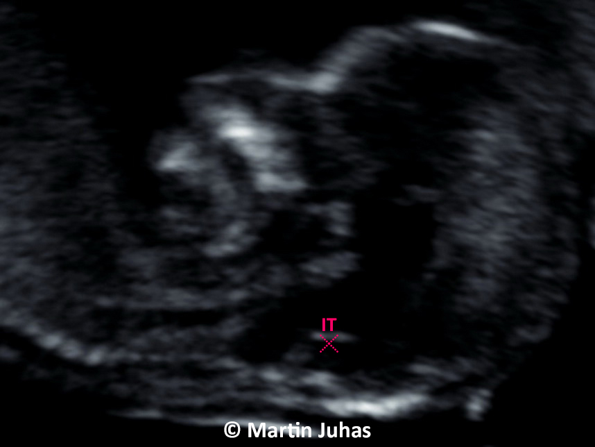
Is it possible to determine schistorrhachis early?
Recently, a new marker, the so-called IT (intracranial translucence), which deals with the risk of the foetus having an opening on the spinal cord – NTD (neural tubal defect) has been added to the morphological examination in the first trimester. This marker shifts the diagnosis of NTD from the period of the second to the first trimester. This examination is based on ultrasound observation of the individual sonomorphological parts of the 4 brain chambers with subsequent evaluation by IT, which is absent with congenital open schistorrhachis.