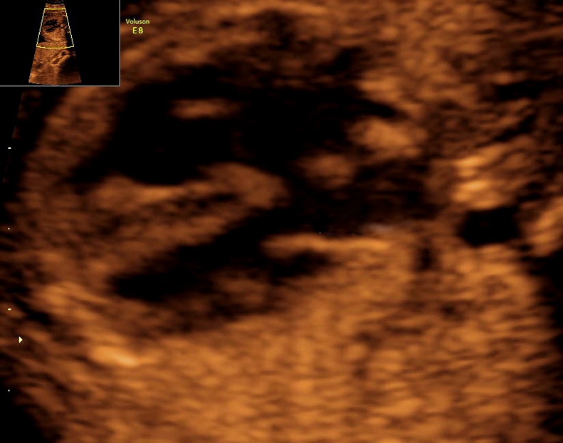
3D/4D ultrasound
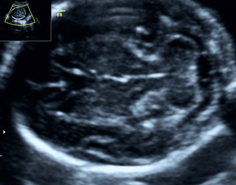
II. trimester
Morphological and genetic ultrasound
Generally
The most important ultrasound examination for excluding congenital developmental birth defects. It evaluates in detail the anatomy and captures the majority of abnormalities of internal organs in the body of the foetus. In the case of the heart and kidneys it’s possible to make determinations also regarding their function. We measure the size of the individual parts of the foetus – typically the diameter and circumference of the head (biparietal diameter - BPD, HC), abdominal circumference (AC) and the length of the leg and arm bones (FL, HL) – by which we assess adequate growth of the foetus. The status of location of the placenta is also evaluated as well as the amount of amniotic fluid, the cervix with possible Doppler metric measurements of the veins of the foetus and mother.
Genetic ultrasound consist of searching for and evaluating defects/anomalies of so-called markers, which often occur in foetuses with chromosomal aberrations (Down, Edwards, Patau Syndrome and others). Such an examination by ultrasound increases the detection of foetuses with Down Syndrome and significantly lowers the necessity of invasive amniocentesis. The aim of genetic ultrasound is to assign to each patient individually – her own risk – for a possible foetus with Down Syndrome and not only general risk derived from the age of the patient or a calculation on the basis of biochemical screening (triple test, double test, serum integrated test...). Detectability of Down Syndrome by genetic ultrasound achieves 91%, whereas with a basic clinical morphological ultrasound this is only 50%.
When?
Morphological ultrasound examination of the foetus is done in the second trimester of pregnancy as a so-called second-trimester ultrasound screening between weeks 19 and 22 of the pregnancy. We perform genetic ultrasound between the 18th to 20th weeks of pregnancy; the given limitation of weeks is linked to the capturing of individual sonograph markers and for potential time possibility of genetic examination either NIPT analysis (link to PRENASCAN) or amniotic fluid – amniocentesis (link to AMC).
How?
We most often carry out the examination with a USG abdominal probe. Before the examination we ask that you empty your urinary bladder and not apply any creams to the abdomen. Always bring all ultrasound findings from your pregnancy as well as other medical documentation with you. Genetic ultrasound is conceived as a second-trimester screening for increasing the capture of foetuses in which chromosomal aberrations are suspected. The idea has found wider application in the USA in particular. Here in Central Europe it is fully substituted by the first-trimester combined test and morphological ultrasound. If, however, a client has not undergone the mentioned screening approach, genetic ultrasound offers the opportunity for negating a positive biochemical screening for Down Syndrome without the need for amniocentesis or NIPT. Here at CFGD we annually negate by genetic ultrasound more than 95% of “triple tests” positive for Down Syndrome. For exact calculation of the risk of chromosomal aberration it is essential that the patient bring the result of a biochemical screening with her.
What is it for?
Basic morphological ultrasound is a standard anatomical examination of the foetus (SSUM recommendation for prenatal examinationand according to these recommendations it is done in the majority of primary gynecological clinics. This standard ultrasound examination is therefore useful, but with respect to its simplicity it has notably specific limitations for the detection of congenital defects.
Expanded morphological ultrasound, which we perform at CFGD, is supplemented compared to basic morphological ultrasound by these examinations:
- detection of minor markers for chromosomal aberrations
- examination of the heart beyond the framework of four-chamber projection
- expanded neurosonogram of individual parts of the foetal brain
- colour, pulsed and tissue Doppler metric measurement
- application of 3D/4D ultrasound modalities
- syndromological detection
- Practice guidelines for performance of the routine mid-trimester fetal ultrasound scan
- ISUOG Practice Guidelines (updated): sonographic screening examination of the fetal heart
- Sonographic examination of the fetal central nervous system: guidelines for performing the ‘basic examination’ and the ‘fetal neurosonogram’
- Screening for chromosomal defects (Nicolaides KH, Screening for chromosomal defects, editorial. Ultrasound Obstet. Gynecol. 2003 Apr;21:313-21.)
- Genetic sonography – the historical and clinical role of fetal echocardiography
- ACR–ACOG–AIUM–SMFM–SRU PRACTICE PARAMETER FOR THE PERFORMANCE OF STANDARD DIAGNOSTIC OBSTETRICAL ULTRASOUND
- Content of a complete second trimester obstetrical ultrasound