Gynecology ambulatory office

Screening for uterine cancer
How does early detection help?
Thanks to the preventive diagnosis with the expert 2D and 3D/4D ultrasound examinations and with subsequent possibility of verification of the actual abnormal ultrasound findings by cytological examinations of the uterine mucosa in the form of pipette collection (TU VABRA), early cervical cancer detection is increased by over 30%. At the same time, it is also important to note the significant impact of this procedure on the overall decrease in curettage – “scraping” in women by more than 90%! This decline can be easily interpreted numerically by saying that almost every third woman in Slovakia has at least once in her life undergone curettage. And yet the statistical rate of endometrial cancer detection remains unchanged over the last 20-30 years. Zároveň treba podotknúť významný dopat uvedeného postupu na celkový pokles kyretáží - “výškrabov” u žien o viac ako 90%! Tento pokles sa dá číselne jednoducho interpretovať tým, že na Slovensku temer každá tretia žena aspoň raz v živote absolvovala kyretáž. Ibaže štatistická miera detekčnosti karcinómu endometria ostáva bez zmeny za posledných 20-30 rokov.
What is the Expert 2D and 3D/4D Ultrasound Examination?
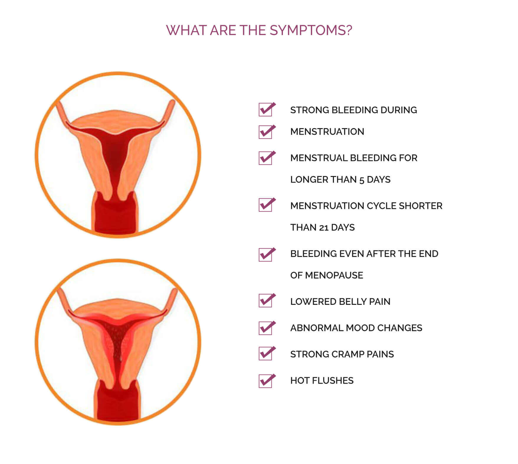
In modern oncogynecology, the basic imaging method is expert ultrasound which has a greater precision than other imaging methods such as CT (computed tomography) or MRI (Magnetic Resonance Imaging). Abnormal bleeding is a common problem in patients, especially during and after menopause. Women with this symptom should be examined immediately by the expert 2D and 3D/4D ultrasound examination, focusing primarily on the uterine mucosa.
- in the dynamic of the examination and imaging in three dimensions of the given organ
- in the possibility of assessing the mobility of an organ, painfulness, movement of fluid content and elastography
- in the combination of sonomorphological and flows examination with the assessment of vascular mucosal architecture and body of the uterus with IETA scoring
- in the most imaging of the uterus and its mucosa
- in the complete absence of ionizing radiation
What is TU VABRA?
TU VABRA—Targeted Ultrasound Vacuum AbraAbrasion is a cytology-bioptic examination of uterine mucosal cells. The catheter aspirator receives a sample of the uterine mucosa that is sent to the histo-cytological examination. Endometrial sampling is a cost-effective method for the first line of diagnostics for abnormal uterine bleeding. The patented configuration of the multiport tip using the TU VABRA pipette is designed to provide a sample from the surface of the uterine mucosa, which improves the detection of focal pathology and minimizes false-negative results.
- fast, effective and painless method
- effective method of the first line of diagnosis
- the silicone, flexible pipette of the TU VABRA cannula eliminates the possibility of cervical trauma
- the 3mm cannula profile facilitates insertion and provides access through a significantly narrowed uterine cervix
- dilation – expansion of the cervix canal – is not required
- anaesthesia is not required, TU VABRA is therefore not associated with a lot of complications in performance, as with curettage
When should you undergo TU VABRA?
Targeted ultrasound biopsy of the uterine mucosa is recommended for abnormal vaginal bleeding during menstrual and especially during menopausal periods, abnormal ultrasound findings and infertility. With this procedure, we are able to capture and diagnose precancerous and cancerous changes on the uterine mucosa or possible infertility in a timely manner, as well as evaluate the mucosa at a certain stage of the menstrual cycle.